Инноваци - Шинэ Санаа, Шинэ Нээлт, 2008, 2(5-2)
Expression of HDAC-1 on the patients with gallbladder cholangiocarcinomas and Intrahepatic cholangiocarcinomas
( Судалгааны өгүүлэл )
Background: Increasing evidence points to a link between histone deacetylases (HDACs) and tumorogenesis. Although several HDAC inhibitors have been tested in clinical trials for cancer therapies, the mechanisms of HDAC activation in tumors remain unknown.
METHODS
Material and Method of HDAC1 on gallbladder carcinomas: The expression of HDAC protein is studied by immunohistochemistry in 31 specimens of gallbladder carcinoma. Formalin fixed and paraffin embedded specimens were obtained from 35 and 31 consecutive patients. Who had an undergone surgical treatment between 1994 and 2003 at the Department of Digestive Intestinal Surgery, Institute of Health biosciences, the University of Tokushima Graduate School.
Antibody: HDAC1 (C-19): sc-6298 goat polyclonal antibody [Santa Cruz] Assessment of HDAC1 staining section tissue: In this cases was assigned the following scores: 0 point: <10%; 1+ point, 10-50%; 2+ point, <50% positive cells. For statistical analysis, the cutoff values for positive expression 10% than more for HDAC1.
RESULTS
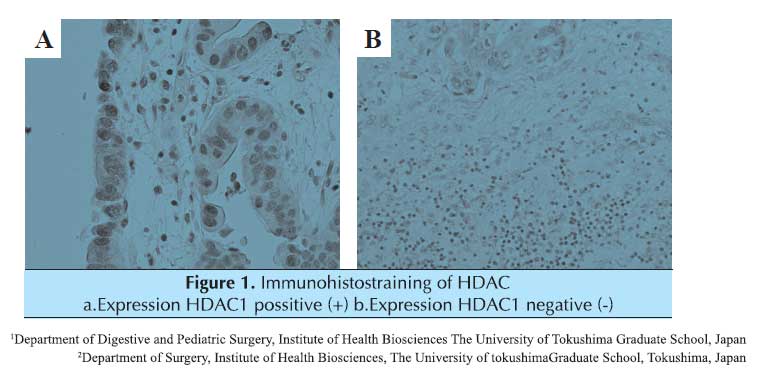 HDAC1(+) positive expression was 20 (64.5%) of 31 cases. HDACl(-) negative expression was 11 (35.5%) of 31 cases.
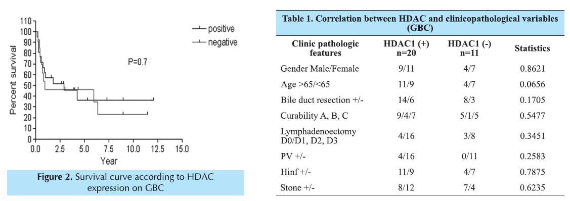
METHODS
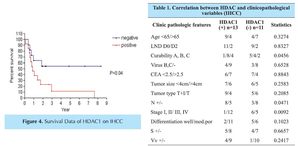
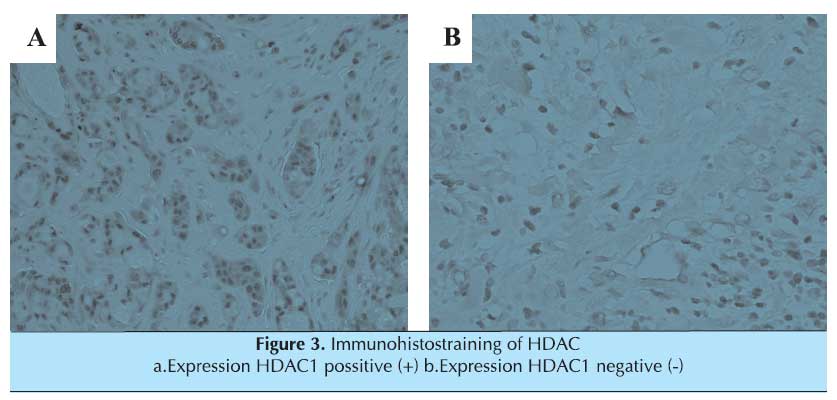
Material and Method of HDAC1 on Intrahepatic cholangiocarcinomas: The expression of HDAC protein is studied by immunohistochemistry in 24 specimens of intrahepatic cholangio carcinoma. Formalin fixed and paraffin embedded specimens were obtained 24 consecutive patients. Who had an undergone surgical treatment between 1994 and 2006 at the Department of Digestive Intestinal Surgery, Institute of Health biosciences, the University of Tokushima Graduate School. Survival data were available in 24 patients. The follow-up period ranged from 1-90 months. All patients alive completed a minimum follow-up of 1 month.
Antibody: HDAC1 (C-19): sc-6298 goat polyclonal antibody [Santa Cruz] Assessment of HDAC 1 staining section tissue: In this cases was assigned the following scores: 0 point: <10%; 1+, 10-50%; 2+,<50% positive cells. For statistical analysis, the cutoff values for positive expression 10% than more for HDAC1.
RESULTS
The positive staining was located in the cytoplasm or nuclear. There was stained HDAC1 (+) expression 13 (54.1%) cases and including 3 strongly positive cases and no stained HDAC1 (-) expression 11 (45.9%) cases.
Ном зүй
1. Claudia A et al. \\\"Histone deacetylase-1 and -3 protein expression in human breast cancer: a tissue microarray analysis\\\" Breast Cancer Research and Treatment (2005) 90: 15-23
2. Hee Jin Chang et al. \\\"Bax, a predictive marker for therapeutic response to preoperative chemoradiotherapy in
patients with rectal carcinoma\\\" Human Pathology (2005)36, 364-371 3. N Hajji et al. \\\"Combinatorial action of the HDAC inhibitor trichostatin A and etoposide induces caspase-mediated AIF-dependent apoptotic cell death in non-small cell lungcarcinoma cells\\\" Oncogene (2007), 1-11
4. Sireerat Pluemsampant et al. \\\"Protein kinase CK2 is a key activator of histone deacetylase in hypoxia-associated tumors\\\" 10.1002/ Infl Journal of Cancer. 23094
Нийтлэлийн нээгдсэн тоо: 509
Судлаачдын бусад өгүүлэл